"Wretched beasties moving about very nimbly"
This is what Anton van Leeuwenhoek said about the creatures he saw in his 1670s microscope. The "Father of Microbiology" made over 400 different types of microscopes and discovered bacteria and spermatozoa, among other things.... among many, many other things. All these "beasties" live in enchanting world and can sometimes look very cute and photographers keep discovering new angles and frontiers of their microcosm.
The "Fairy Fly" wasp (left) and some iridescent part of the aptly-named "Jewel Beetle":
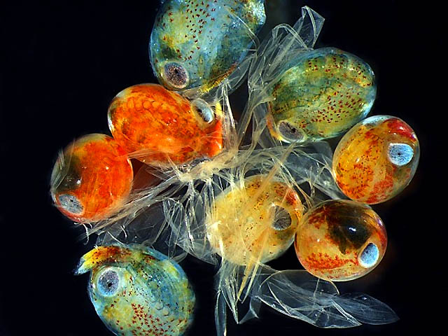
Lobster Eggs:
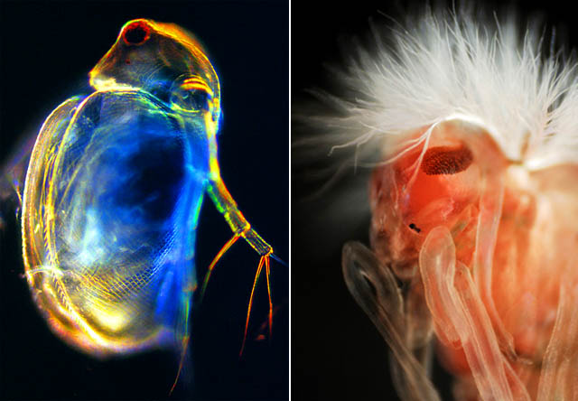
Left: Live Daphnia... and the portrait of a fly (with a swanky hairdo):
Larva of Brachiolaria looks like a wannabe squid:
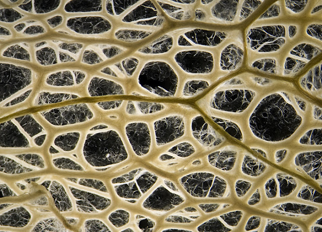
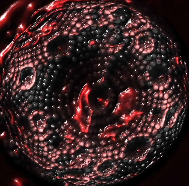
Peeking inside the wild cucumber (Echinocystis lobata) -
The eye of the honeybee (left). And the flatworm on the right seems to have a face. Pretty morose kind of face:
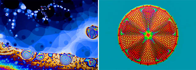
Soap bubbles on the left look somewhat like Mandelbrot set, and Actinoptychus heliopelta on the right looks remarkably like a mandala:
------------
Best Microscope Photos, according to Nikon Small World and National Geographic
Sponsored by Nikon, the annual
Small World Contest showcases "the beauty and complexity of life as seen through the light microscope." See all the winners and honorable mentions
here, and also
presentation on National Geographic.
Nanotube Factory:
"Nanotubes are elongated, hollow cylinders of carbon atoms, just 1/50,000 the width of a human hair... Sometimes, the heated mass of nanotubes grows like a bulb in the spring."
 Photograph by Paul Marshall/National Research Council Canada /courtesy of Nikon Small World
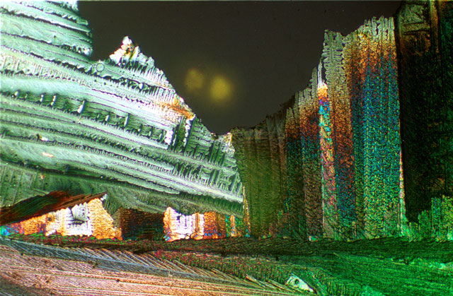
Drug Landscape:
The antibiotic powder mitomycin - "viewed through polarizing filters, the drug gave off colors that reveal its complex crystal structure."
A Chick Embryo:
 Photograph by Thomas Pais de Azevedo of Lisbon, Portugal/photo courtesy of Nikon Small World
And if you (like us) can't get enough of microscope photography, then this link is for you: the gallery of photomicrographs by year dating back to 1977! -
Click Here.
Another great set of microscope photos is
here - "Zoomified", by Tracy E. Anderson.
------------
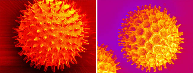
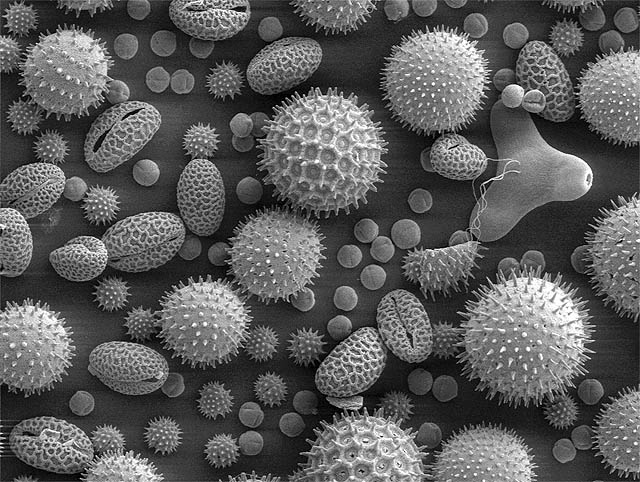
Images of Pollen
Rippel Electron Microscope Facility shows images of
Ipomea purpurea (Heavenly blue morning glory) pollen - see
here, including three-dimensional ones.
Truly a heavenly mix:
------------
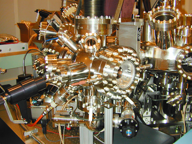
Extreme Zoom! Nano-scale Explorations
A scanning tunneling microscope (based on the concept of
quantum tunneling) and Field Ion Microscopy Systems allow us to see individual atoms (
here is a good article about it) The first images of atomic structures were published as far back as 1951.
Field Ion Microscope also allows to sharpen metal tips (usually tungsten needles) to the ultimate degree - producing The Sharpest Manmade Thing:
What you see is a tip of that needle: the small round objects are individual atoms.
For those who love retro hardware: Soviet early-60s Electronic Microscope (featuring an electron gun) -
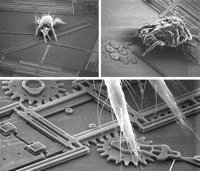
Bugs on Chips: Microelectromechanical systems
MEMS are almost nano-scale micromachines... Here microscope photography helps to establish the size of these machines, compared to less than 1mm mite, or a mite's leg:
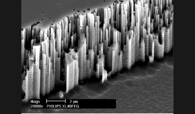
Is this a microscopic Hong Kong? -
No, this is so-called Bosch process plasma etchings - image done for The 50th International Conference on Electron, Ion and Photon Beam Technology, see previous entries
here.
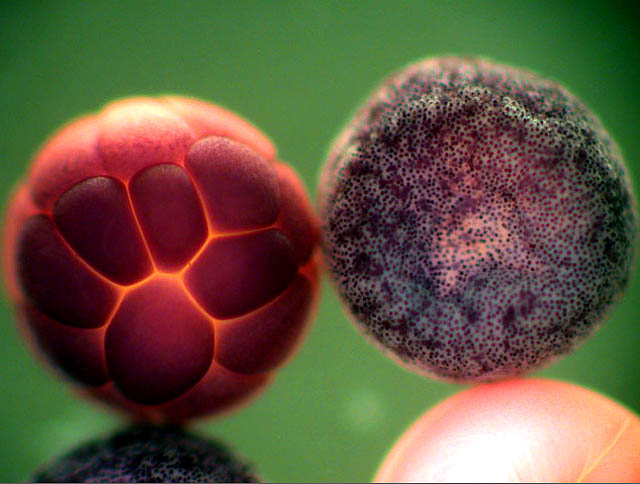
Frog Embryos:
Microscopic frost accumulating on a blade of grass -
------------


 Reply With Quote


Social Networking Bookmarks