Peering into the micro world
A team of University of Michigan researchers has recently created a set of electron microscope images of carbon nanotube structures depicting images of U.S. President-elect Barack Obama. John Hart, leader of the research team says it wasn't a political statement, but an attempt to draw attention to what is possible these days with nanotechnology, and imaging at the very small scale. I'll take him up on this invitation and share with you some other images of very tiny things in our world. For visualizing the scale, most measurements below are in microns - one micron is a millionth of a meter - human hair is approximately 100 microns thick. (
32 photos total)

Images of U.S. President-elect Barack Obama, each made with approximately 150 million tiny carbon nanotubes, are photographed using an electron microscope by University of Michigan Mechanical Engineering Department in this image released to Reuters November 10, 2008. The image, based on an original drawing by Shepard Fairey, is just wider than 500 microns and is made of approximately 150 million tiny carbon nanotubes, which is about the number of Americans who voted on November 4, according to John Hart at University of Michigan. (REUTERS/John Hart, Sameh Tawfick, Michael De Volder, and Will Walker/University of Michigan/Handout)
 2
2
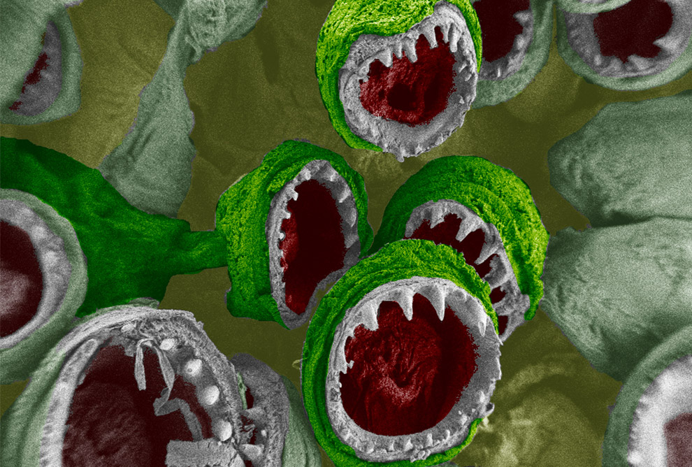
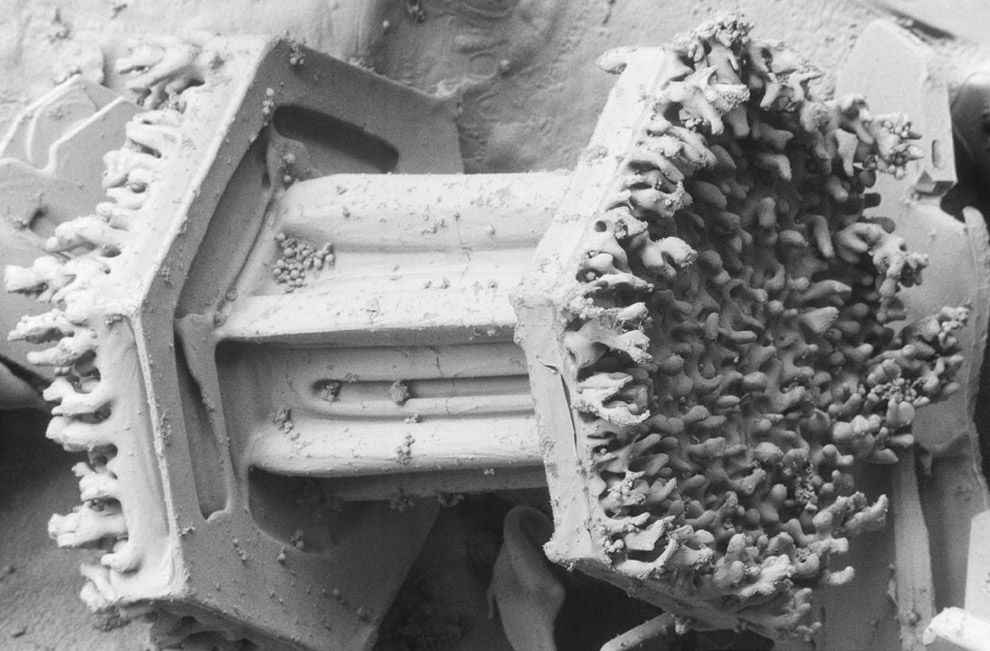
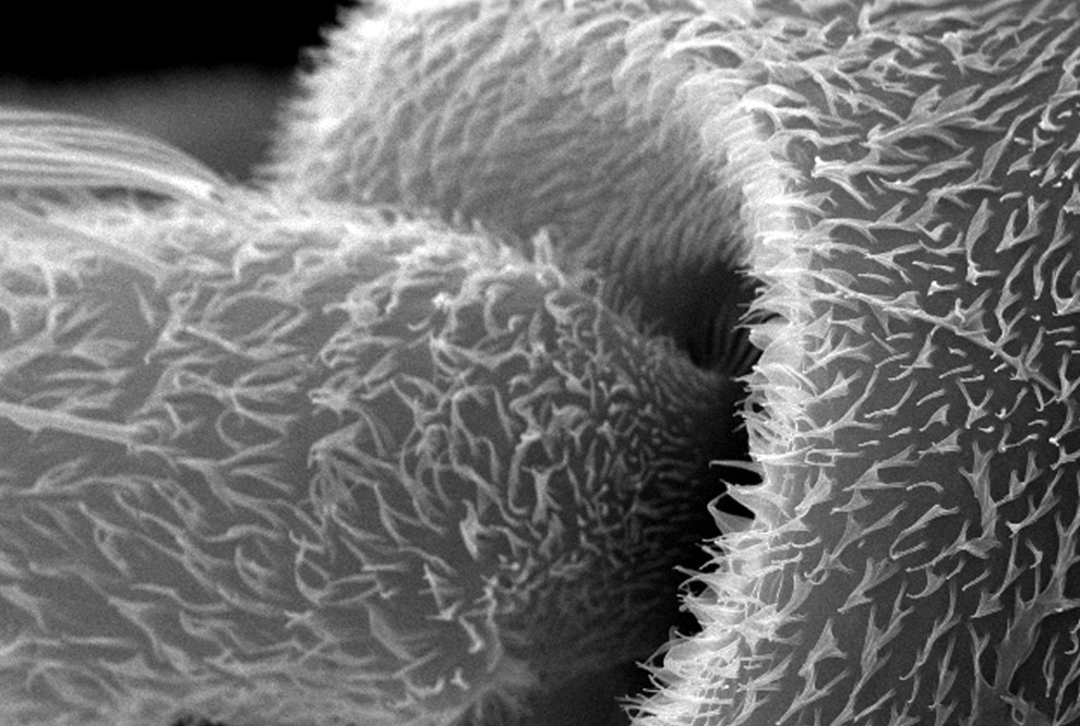
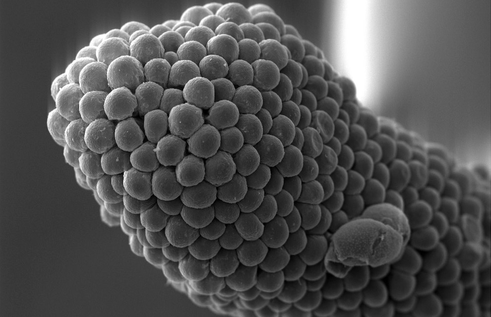
Squid Suckers, winner of Honorable Mention in the 2008 International Science and Engineering Visualization Awards. Loligo pealei squids have eight arms and two tentacles, all of which are coated with suction-cups, lined with fangs composed of chitin. These tiny suckers, whose diameters are around 400 microns, ultimately allow the half-meter-long squid to get a solid grip on its environment. (Courtesy of Jessica D. Schiffman and Caroline L. Schauer; Drexel University)
#
 3
3
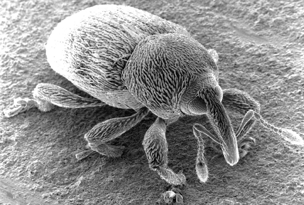
Electron micrograph close-up of a weevil (Curculionidae family) - its snout is just over 100 microns wide. (Dartmouth Electron Microscope Facility/Dartmouth College)
#
 4
4
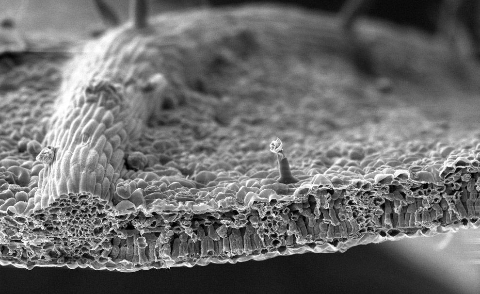
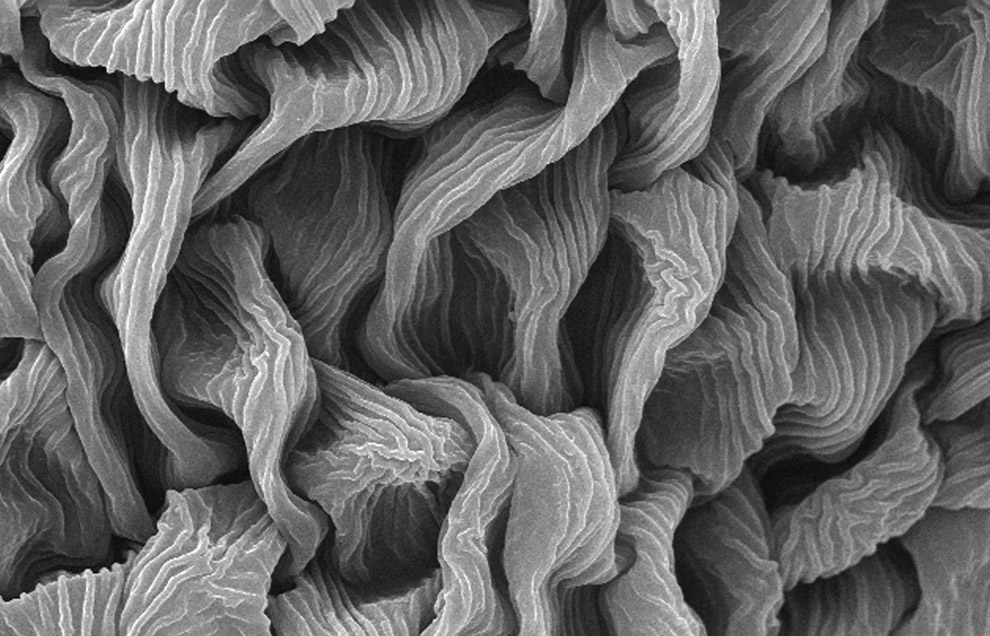
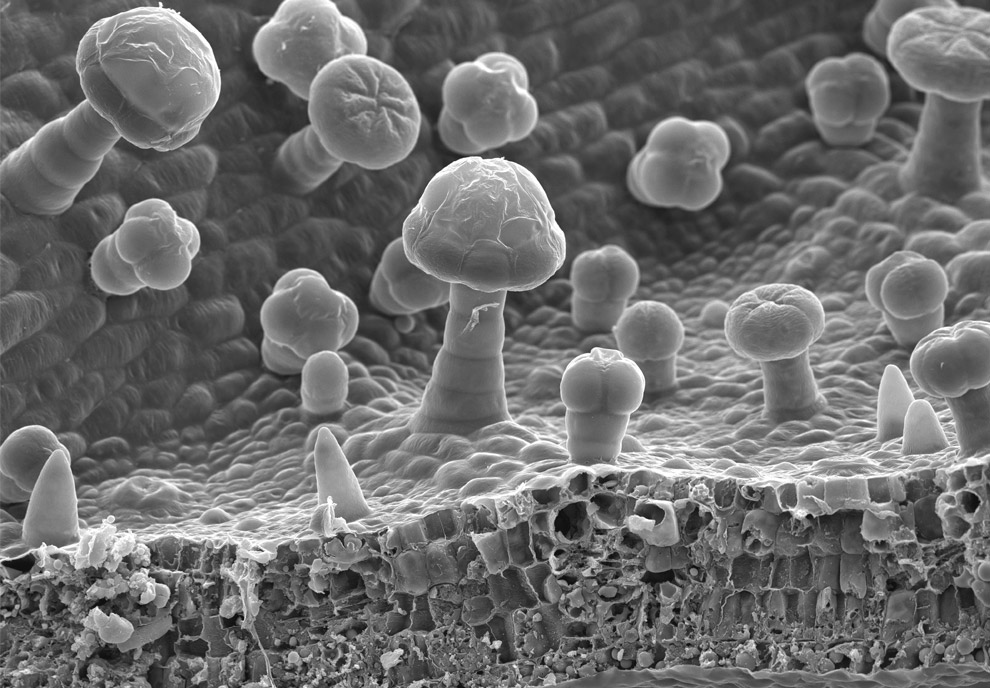
Scanning electron microscope image of a leaf from a Black Walnut tree. Image shows a cross-section of a cut leaf, itsupper epidermal layer, mesophyll layer with palisade cells and vascular bundles, and lower epidermal layer. The protrusion at center is just over 50 microns tall. (Dartmouth Electron Microscope Facility/Dartmouth College)
#
 5
5
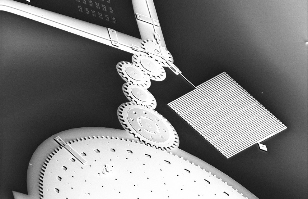
A Microelectromechanical system (MEMS) gear train manufactured on silicon. The larger gear at center is about 80 microns wide. (Institute of Micromachine and Microfabrication Research at Simon Fraser University)
#
 6
6
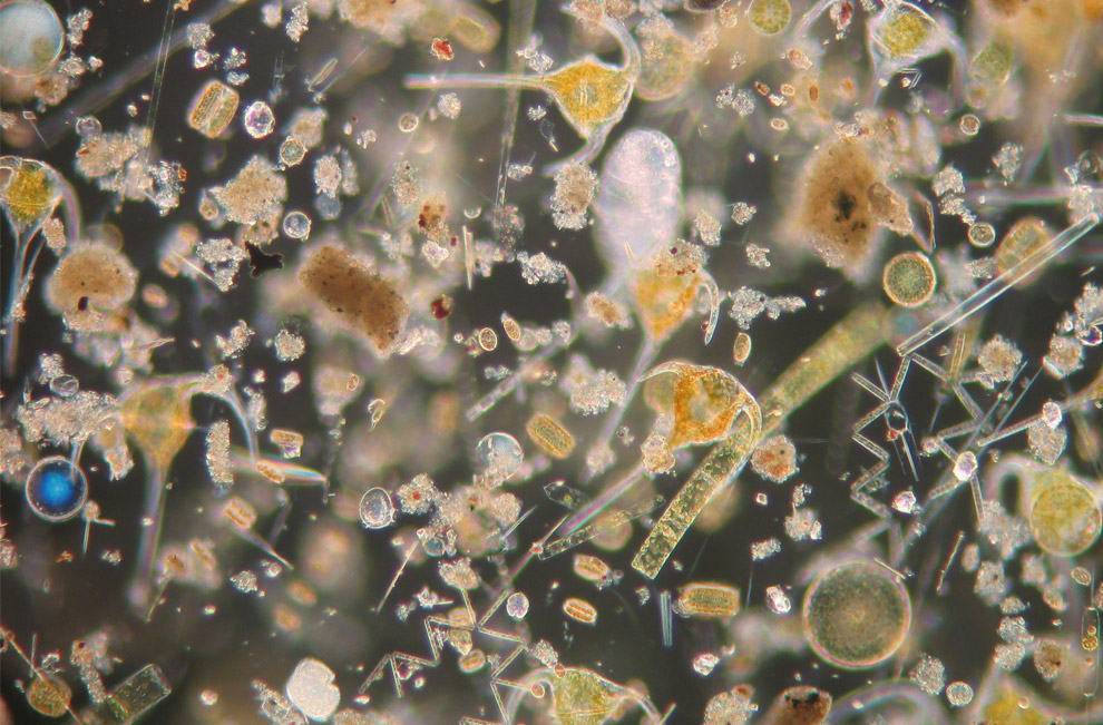
Microalgae seen under the microscope: CO2-feeders in the ocean. (A. Stuhr/ IFM-GEOMAR)
#
 7
7
Rime on a columnar snow crystal. Contact between the snow crystal and the supercooled droplets in the air resulted in freezing of the liquid droplets onto the surface of the crystal. Observations of snow crystals clearly show cloud droplets measuring up to 50 microns on the surface of the crystal. (Agricultural Research Service, United States Department of Agriculture)
#
 8
8
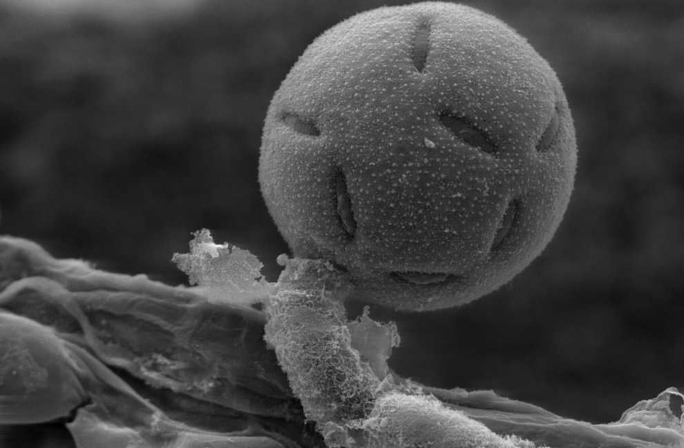
Pollen from a variety of common plants: sunflower, morning glory, hollyhock, lily, primrose and caster bean. The largest one at center is nearly 100 microns wide. (Dartmouth Electron Microscope Facility/Dartmouth College)
#
 9
9
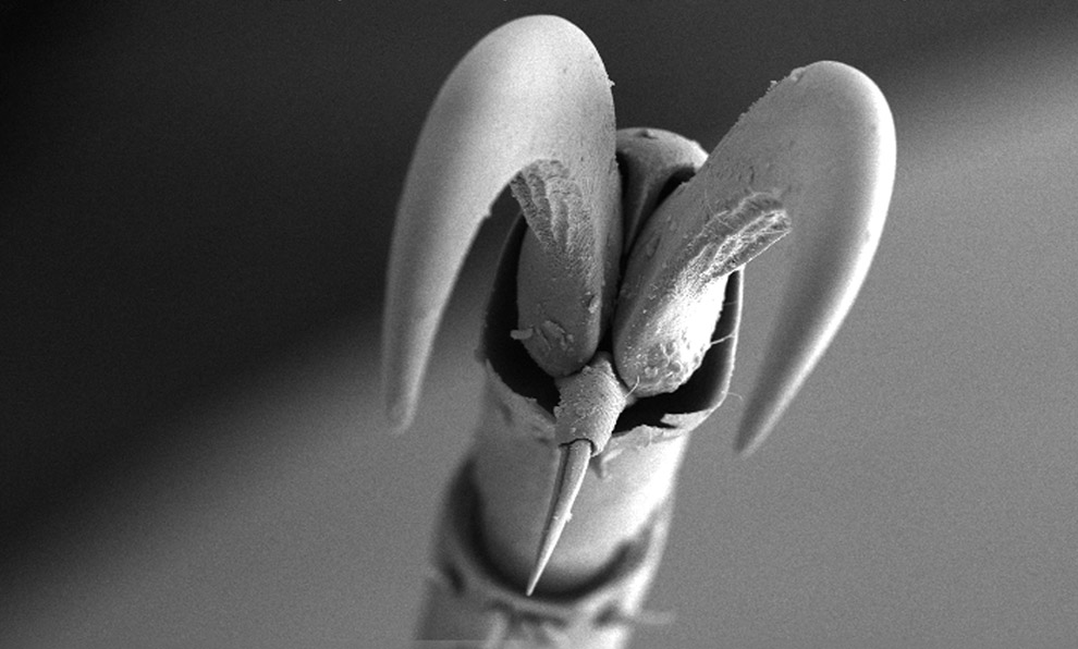
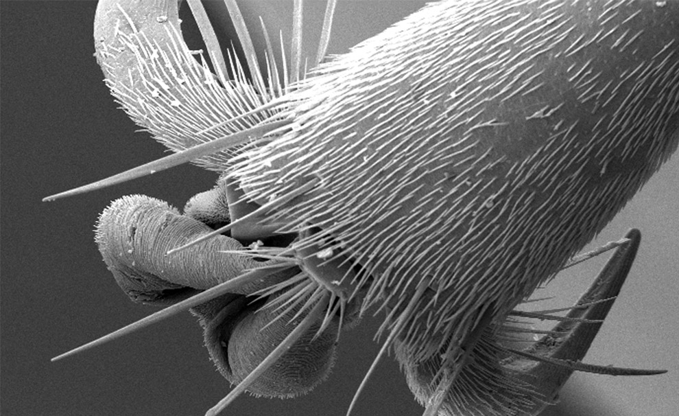
At a magnification of 94X, this is a scanning electron micrograph view of the distal clawed tip of an adult figeater beetle's leg. The insect leg is comprised of a variable number of segments, incliuding the pretarsus, seen here with a claw and spiked empodium. (CDC/Janice Carr)
#
 10
10
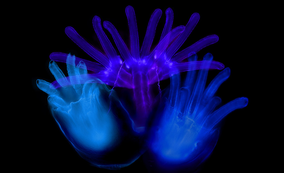
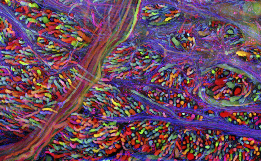
n this Harvard University photograph released October 8th, 2008, brain cells of a laboratory mouse are shown glowing with multicolor fluorescent proteins at Harvard University in Cambridge, Mass. The Nobel prize in chemistry was awarded to two Americans and a U.S.-based Japanese scientist for research on a glowing jellyfish protein that revolutionized the ability to study disease and normal development in living organisms. (AP Photo/Harvard University, Livett-Weissman-Sanes-Lichtman)
#
 11
11
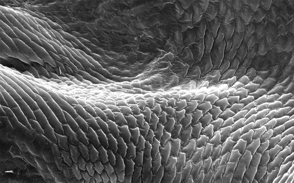
Magnified 598x, this scanning electron micrograph depicts an enlarged view of the chitinous, exoskeletal surface of a male louse, Pediculus humanus var. corporis. In this particular view, the exoskeleton appears to be composed of interlocking plates. (CDC/Janice Carr)
#
 12
12
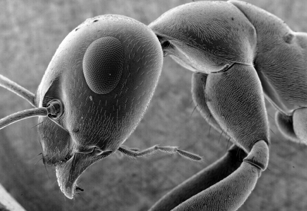
Scanning electron microscope image of an ant. Its eye is approximately 300 microns wide. (Dartmouth Electron Microscope Facility/Dartmouth College)
#
 13
13
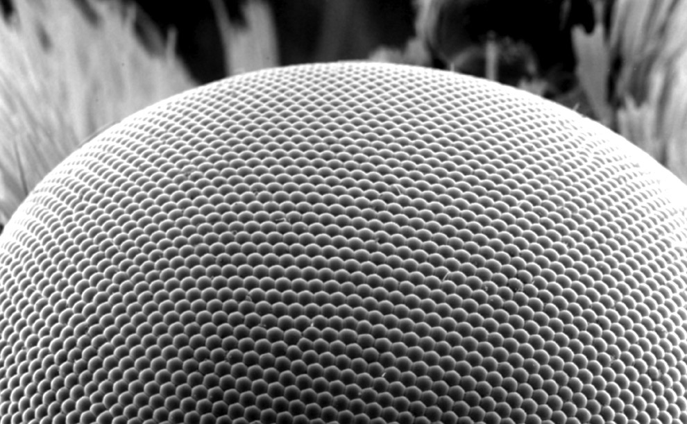
Scanning electron microscope image of the compound eye of a noctuid moth. Each facet of the eye (ommatidium) is approximately 25 microns wide. (Dartmouth Electron Microscope Facility/Dartmouth College)
#
 14
14
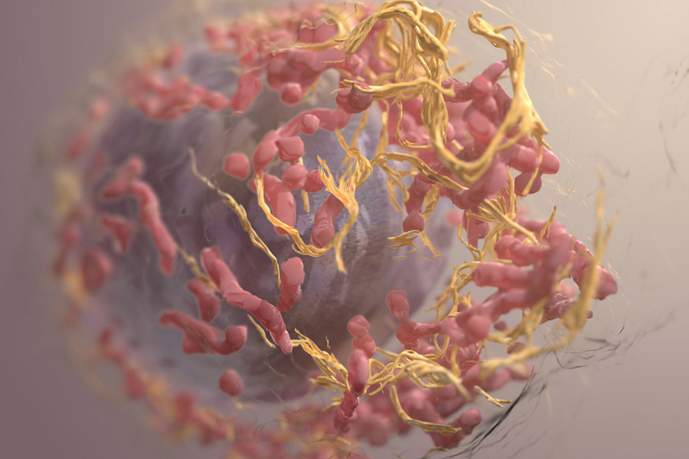
Winner of an Honorable Mention in Illustration in the 2008 International Science and Engineering Visualization Awards, this is a rendered 3D image of a melanoma cell using data obtained using ion abrasion scanning electron microscopy, a novel approach for imaging mammalian cells at nanometer resolution. (Donald Bliss and Sriram Subramaniam; National Library of Medicine, NIH)
#
 15
15
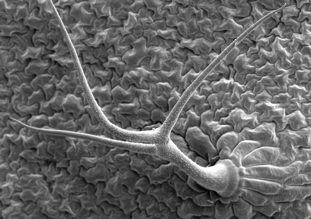
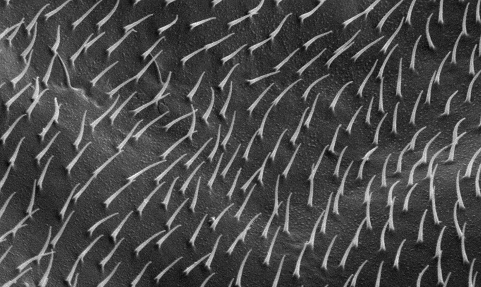
Scanning electron microscope image of lower surface of Arabidopsis thaliana leaf, showing a trichome - an outgrowth, or "leaf hair" that grows out of specialized epidermal cells. (Dartmouth Electron Microscope Facility/Dartmouth College)
#
 16
16
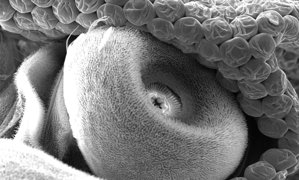
Scanning electron microscope image of a pyralidae moth, a side view of its head and curled proboscis. Its eye is about 800 microns wide. (Dartmouth Electron Microscope Facility/Dartmouth College)
#
 17
17
Under a magnification of 1438x, this scanning electron micrograph reveals some of the ultrastructural details seen on the surface of a "crimson clover", Trifolium incarnatum flower petal. (CDC/Janice Carr)
#
 18
18
The anterior spiracles (respiratory openings) of a fruit fly larvae magnified 1500x. (Albert Tousson and Tomek Szul; Department of Cell Biology The University of Alabama at Birmingham)
#
 19
19
Under high magnification of 5653x, this scanning electron micrograph depicts the surface of an unidentified insect's compound eye, revealing photoreceptor cells, support cells and pigment cells that make up the repeating hexagonally-shaped units of a compund eye known as "ommatidia". (CDC/Janice Carr)
#
 20
20
This scanning electron micrograph shows the exoskeletal morphology found on one of the six legs of an unidentified hornet found in the suburbs of Decatur, Georgia. Under a magnification of 87X, what this SEM reveals is the anatomical configuration of what is termed the leg's "tarsal chain", which comprises the tarsus, and pretarsus or claw. (CDC/Janice Carr)
#
 21
21
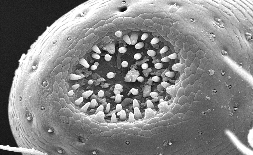
At a magnification of 765X, this scanning electron micrograph reveals morphologic details found at the tip of this adult figeater beetle's maxillary galea, which due to its shape, was given this Latin name for "helmet". The galea is located just medial to another, more prominent maxillary appendage, the palpus. Note the concave configuration at the distal tip of the galea, and how there are numerous pointed protuberances inside this concavity, which are most probably sensorial in nature. (CDC/Janice Carr)
#
 22
22
Scanning electron microscope image of Heliotropium (marine heliotrope) lower leaf surface, showing trichomes and a few stomata. The large trichome at bottom is approximately 50 microns wide at its base. (Dartmouth Electron Microscope Facility/Dartmouth College)
#
 23
23
Description: At a magnification of 1504x, this scanning electron micrograph shows features of an Anopheles dirus mosquito's antennae. In this particular view, only the first two (of three) segments of the left antenna are visible. Covered with sensorial "hairs", which aren't really hairs at all, but exoskeletal chitinous extensions, known as "setae", they provide feedback to the mosquito as to chemical, thermal, and tactile changes in its environment. (CDC/Janice Carr)
#
 24
24
n this Harvard University photograph released October 8th, 2008, brain cells of a laboratory mouse are shown glowing with multicolor fluorescent proteins at Harvard University in Cambridge, Mass. The Nobel prize in chemistry was awarded to two Americans and a U.S.-based Japanese scientist for research on a glowing jellyfish protein that revolutionized the ability to study disease and normal development in living organisms. (AP Photo/Harvard University, Livett-Weissman-Sanes-Lichtman)
#
 25
25
Under a low magnification of 58X this scanning electron micrograph shows some of the exoskeletal morphologic characteristics displayed on the head region of an unidentified beetle. What appears to be hairs are actually sensorial organs known as "setae", which provide the organism with information about its environment including changes in temperature, wind direction, and chemical queues, i.e., pheromones. (CDC/Janice Carr)
#
 26
26
This scanning electron micrograph shows the "scape", or the first segment of an unidentified mosquito's left antenna magnified 500X. Note that the central region of the scape is concave, where the second segment of the antenna, known as the "pedicle" will interlock. The grape-like ommatidia surrounding the scape are the functional units of its compound eyes. (CDC/Janice Carr)
#
 27
27
A polllen grain on perched on the anther of a Penta lanceolata flower. The grain is about 40 microns wide. (Dartmouth Electron Microscope Facility/Dartmouth College)
#
 28
28
Scanning electron microscope image of the stigma of a Penta lanceolata flower, approximately 140 microns wide. (Dartmouth Electron Microscope Facility/Dartmouth College)
#
 29
29
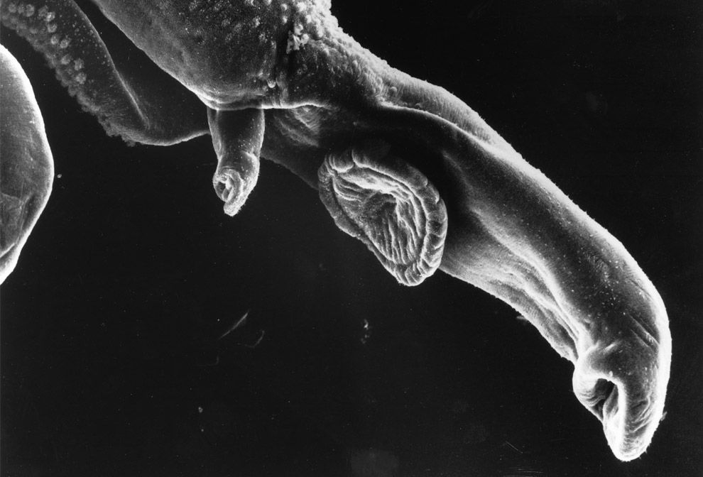
This image is of a schistosome parasite. It enters the body through the skin of persons coming in contact with infested waters. The adult worm lives in the veins of its host. The parasite is magnified x256 in this photograph. (National Cancer Institute / Bruce Wetzel, Harry Schaefer)
#
 30
30
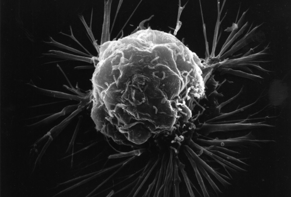
Pictured is a breast cancer cell, photographed by a scanning electron microscope. This picture shows the overall shape of the cell's surface at a very high magnification. Cancer cells are best identified by internal details, but research with a scanning electron microscope can show how cells respond in changing environments and can show mapping distribution of binding sites of hormones and other biological molecules. (National Cancer Institute)
#
 31
31
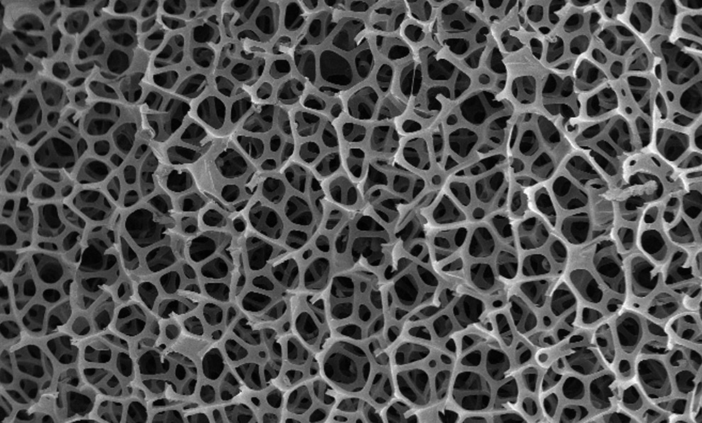
Under a low magnification of 23x, this 2007 scanning electron micrograph depicts the fibrous configuration of a dry macrofoam sponge swab. (CDC/Janice Carr)
#
 32
32
Scanning electron microscope image of Juglans nigra (Black Walnut tree) lower leaf surface, showing a variety of trichomes. (Dartmouth Electron Microscope Facility/Dartmouth College)
#


Originally Posted by prezzy

 Posting Permissions


 Reply With Quote
Social Networking Bookmarks